Endoscopic Retrograde Cholangiopancreatography (ERCP)
 Is a procedure that combines the use of endoscopy and fluoroscopy to diagnose and treat certain problems of the liver, pancreas and gallbladder.
Endoscopic retrograde cholangiopancreatography (ERCP) is a procedure that combines upper gastrointestinal (GI) endoscopy and x-rays to treat problems of the bile and pancreatic ducts.
What are the bile and pancreatic ducts?
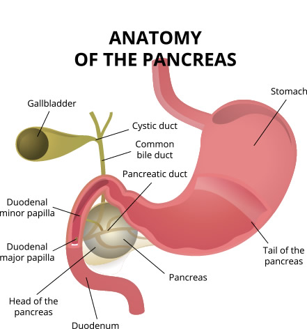
Your bile ducts are tubes that carry bile from your liver to your gallbladder and duodenum. Your pancreatic ducts are tubes that carry pancreatic juice from your pancreas to your duodenum. Small pancreatic ducts empty into the main pancreatic duct. Your common bile duct and main pancreatic duct join before emptying into your duodenum.
We use ERCP to treat problems of the bile and pancreatic ducts. We also use ERCP to diagnose problems of the bile and pancreatic ducts if they expect to treat problems during the procedure. For diagnosis alone, your doctor may use noninvasive tests that do not physically enter the body instead of ERCP. Noninvasive tests such as magnetic resonance cholangiopancreatography (MRCP) a type of magnetic resonance imaging (MRI) are safer and can also diagnose many problems of the bile and pancreatic ducts.
How do I prepare for ERCP?
To prepare for ERCP, talk with your doctor, arrange for a ride home, and follow your doctor’s instructions.
You should talk with your doctor about any allergies and medical conditions you have and all prescribed and over-the-counter medicines, vitamins, and supplements you take, including:
- arthritis medicines
- aspirin or medicines that contain aspirin
- blood thinners
- blood pressure medicines
- diabetes medicines
- nonsteroidal anti-inflammatory drugs (NSAIDs) such as ibuprofen and naproxen
Your doctor may ask you to temporarily stop taking medicines that affect blood clotting or interact with sedatives. You typically receive sedatives during ERCP to help you relax and stay comfortable.
Tell your doctor if you are, or may be, pregnant. If you are pregnant and need ERCP to treat a problem, the doctor performing the procedure may make changes to protect the fetus from x-rays. Research has found that ERCP is generally safe during pregnancy.
How do doctors perform ERCP?
Our doctors specialized training in ERCP perform this procedure at a hospital or an outpatient center. An intravenous (IV) needle will be placed in your arm to provide a sedative. Sedatives help you stay relaxed and comfortable during the procedure. A health care professional will give you a liquid anesthetic to gargle or will spray anesthetic on the back of your throat.
You’ll be asked to lie on an examination table. Your doctor will carefully feed the endoscope down your esophagus, through your stomach, and into your duodenum. A small camera mounted on the endoscope will send a video image to a monitor. The endoscope pumps air into your stomach and duodenum, making them easier to see.
During ERCP
The doctor locates the opening where the bile and pancreatic ducts empty into the duodenum slides a thin, flexible tube called a catheter through the endoscope and into the ducts injects a special dye into the ducts through the catheter to make the ducts more visible on x-rays uses a type of x-ray imaging, called fluoroscopy, to examine the ducts and look for narrowed areas or blockages. Instruments are used through the endoscopy to:
- open blocked or narrowed ducts.
- break up or remove stones.
- perform a biopsy or remove tumors in the ducts.
- insert stents—tiny tubes that a doctor leaves in narrowed ducts to hold them open.
- Your doctor may also insert temporary stents to stop bile leaks that can occur after gallbladder surgery.
- The procedure most often takes between 1 and 2 hours.

 Meet Dr. Mark Murray
 Meet Dr. Eric M. Hill
 Meet Dr. kevin Ho